Control Systems
Humans and other highly evolved animals have developed two main systems for coordinating and synchronizing the functions of their millions of individual cells. The nervous system works rapidly by transmitting electrochemical impulses. The endocrine system is a slower system of control; it works by releasing chemical signals into the circulation. In addition to coordinating essential bodily functions, these two control systems allow the animal to react to both its external and internal environments.
The Nervous System
The nervous system functions by the almost instantaneous transmission of electrochemical signals. The means of transmission are highly specialized cells known as neurons, which are the functional unit of the nervous system.
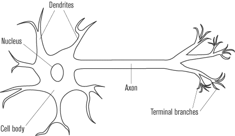
The neuron is an elongated cell that usually consists of three main parts: the dendrites, the cell body, and the axon. The typical neuron contains many dendrites, which have the appearance of thin branches extending from the cell body. The cell body of the neuron contains the nucleus and organelles of the cell. The axon, which can sometimes be thousands of times longer than the rest of the neuron, is a single, long projection extending from the cell body. The axon usually ends in several small branches known as the axon terminals. Neurons are often connected in chains and networks, yet they never actually come in contact with one another. The axon terminals of one neuron is separated from the dendrites of an adjacent neuron by a small gap known as a synapse.
The electrical impulse moving through a neuron begins in the dendrites. From there, it passes through the cell body and then travels along the axon. The impulse always follows the same path from dendrite to cell body to axon. When the electrical impulse reaches the synapse at the end of the axon, it causes the release of specialized chemicals known as neurotransmitters. These neurotransmitters carry the signal across the synapse to the dendrites of the next neuron, starting the process again in the next cell.
The Resting Potential
To understand the nature of the electrical impulse that travels along the neuron, it is necessary to look at the changes that occur in a neuron between when it is at rest and when it is carrying an impulse. When there is no impulse traveling through a neuron, the cell is at its resting potential and the inside of the cell contains a negative charge in relation to the outside.
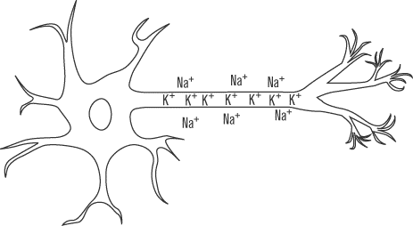
Maintaining a negative charge inside the cell is an active process
that requires energy. The cell membrane of the neuron contains a
protein called Na+ K+ ATPase that uses the
energy provided by one molecule of ATP to pump three positively
charged sodium atoms (Na+)out of the cell, while simultaneously taking into the cell two
positively charged potassium ions (K+).The sodium-potassium pump builds up a high concentration of sodium
ions outside the cell and an excess of potassium ions inside the cell.
These ions naturally want to diffuse across the membrane to regularize
the distribution. However, one of the special properties of
phospholipid cell membranes is that they bar passage to ions unless
there is a special protein channel that allows a particular ion in or
out. No such channel exists for the sodium that is built up outside
the cell, though there are potassium leak channels that allow
some of the potassium ions to flow out of the cell. The difference in
ion concentrations creates a net potential difference across the cell
membrane of approximately –70mV (millivolts), which is the value of the resting potential.
The Action Potential
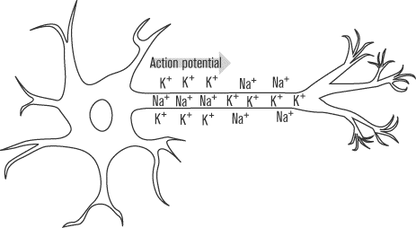
While most cells have some sort of resting potential from the movement
of ions across their membranes, neurons are among only a few types of
cells that can also form an action potential. The action potential is
the electrochemical impulse that can travel along the neuron. In
addition to the Na+K+ ATPase and potassium
leak channel proteins, the neuron membrane contains voltage-gated
proteins. These proteins respond to changes in the membrane
potential by opening to allow certain ions to cross that would not
normally be able to do so. The neuron contains both voltage-gated
sodium channels and voltage-gated potassium channels, which open under
different circumstances.
The action potential begins when chemical signals from another neuron
manage to depolarize, or make less negative, the potential of the cell
membrane in one localized area of the neuron cell membrane, usually in
the dendrites. If the neuron is stimulated enough so that the cell
membrane potential in that area manages to reach as high as
–50 mV (from the resting potential of –70 mV), the
voltage-gated sodium channels in that region of the membrane open up.
The voltage at which the voltage-gated channels open is called the
threshold potential, so the threshold potential in this case is
–50 mV. Since there is a large concentration of positive sodium ions just outside the cell membrane
that have been pumped out by Na+/ K+ ATPase, when the
voltage-gated channels open, these sodium ions follow the concentration gradient and rush into the cell. With the flood of positive ions, the cell continues to depolarize. Eventually the
membrane potential gets as high as +35
mV, at which point the voltage-gated sodium channels close again and
voltage-gated potassium channels reach their threshold and open up.
The positive potassium ions concentrated in the cell now rush out of
the neuron, repolarizing the cell membrane to its negative resting
potential. The membrane potential continues to drop, even beyond
–70mV, until the voltage-gated s
potassium channels close once again at around
–90 mV. With the voltage-gated
proteins closed, the Na+
K+ ATPase and the
potassium leak channels work to restore the membrane potential to its
original polarized state of –70
mV. The whole process takes approximately one millisecond to occur.
Provide the connection between sensory neurons and motor neurons.
The Central Nervous System
The central nervous system consists of the brain and the spinal cord. The spinal cord is a long cylinder of nervous tissue that extends along the vertebral column from the head to the lower back.
Composed of many distinct structures working together to coordinate the body, the brain is a highly complex (and poorly understood) organ. Luckily, you don’t have to “understand” the brain for the SAT II Biology. You just need to know its basic structures and their functions. The brain is made up almost entirely of interneurons.
- The cerebrum is the largest portion of the brain and the seat of consciousness. The cerebrum controls all voluntary movement, sensory perception, speech, memory, and creative thought.
- The cerebellum does not initiate voluntary movement, but it helps fine-tune it. The cerebellum makes sure that movements are coordinated and balanced.
- The brainstem, specifically a portion of it known as the medulla oblongata, is responsible for the control of involuntary functions such as breathing, cardiovascular regulation, and swallowing. The medulla oblongata is absolutely essential for life and processes a great deal of information. The medulla also helps maintain alertness.
- The hypothalamus is responsible for the maintenance of homeostasis. It regulates temperature, controls hunger and thirst, and manages water balance. It also helps generate emotion.
The spinal cord contains all three types of neurons. Axons of motor neurons extend from the spinal column into the peripheral nervous system, while the fibers of sensory neurons merge into the column from the PNS. Interneurons link the motor and sensory neurons, and they make up the majority of the neurons in the spinal column. In addition to the neurons, cells called glial cells are present to provide physical and metabolic support for neurons. The spinal cord serves as a link between the body and the brain, and it can also regulate simple reflexes.
the body and the brain, and it can also regulate simple reflexes.
The brain and spinal cord are bathed in a fluid called the cerebrospinal fluid, which helps to cushion these delicate organs against damage. The cerebrospinal fluid is maintained by the glial cells.
The Peripheral Nervous System
The peripheral nervous system consists of a sensory system that carries information from the senses into the central nervous system from the body and a motor system that branches out from the CNS to targeted organs or muscles. The motor division can be divided into the somatic system and the autonomic system.
The somatic nervous system is responsible for voluntary, or conscious, movement. The neurons only target the skeletal muscles responsible for body movement. All of the neurons in the somatic system release acetylcholine, an excitatory neurotransmitter that causes skeletal muscles to contract. None of the neurons in the somatic nervous system has an inhibitory effect.
The autonomic system controls tissues other than skeletal muscles, including smooth and cardiac muscle, glands, and organs. The system controls processes that an animal does not have voluntary control over, such as the heartbeat, the movements of the digestive tract, and the contraction of the bladder. Autonomic neurons can either excite or inhibit their target muscles or organs. The autonomic nervous system can itself be subdivided into the sympathetic division and parasympathetic division. These two systems act antagonistically and often have opposite effects.
- The sympathetic division prepares the body for emergency situations. It increases the heart rate, dilates the pupils, increases the breathing rate, and diverts blood from the digestive system so that it can be used to oxygenate skeletal muscles that may be needed for action. The sympathetic division also stimulates the medulla of the adrenal glands to release epinephrine and norepinephrine into the bloodstream, hormones that help to reinforce the direct effects of the neurons. Together, the actions of the sympathetic nervous system are often called the “fight or flight” response. The neurotransmitter most often released by sympathetic neurons is norepinephrine.
- The parasympathetic division is most active when the body is at rest. It slows the heart rate, increases digestion, and slows breathing. The effects of the parasympathetic division are sometimes called the “rest and digest” response. The neurotransmitter most often associated with the parasympathetic division of the autonomic nervous system is acetylcholine.
Next to display next topic in the chapter.
Practice Questions
Test Prep Lessons With Video Lessons and Explained MCQ
Large number of solved practice MCQ with explanations. Video Lessons and 10 Fully explained Grand/Full Tests.