The Discovery of Cells
Most cells are too small to be observed with the naked eye. For this reason, even the existence of cells escaped notice until scientists first learned to harness the magnifying power of lenses in the second half of the seventeenth century. At that time a Dutch clothing dealer named Antonie van Leeuwenhoek (1632–1723) fashioned extraordinarily accurate single-lens microscopes. Gazing into the lens of these microscopes, he discovered single-celled organisms, which he called “animalcules” and which, today, we call bacteria and protists.
Englishman Robert Hooke (1635–1703) expanded on Leeuwenhoek’s observations with the newly developed compound microscope, which uses two or more aligned lenses to increase magnification while reducing blurring. When Hooke turned the microscope on a piece of cork, he noticed that the tiny, boxlike compartments of the wood resembled the cells of a monastery. The term “cell” was born.
Microscope and Discovery of Cell
Cell Theory Emerges
As microscope technology improved, scientists were able to study cells in ever-greater detail. Hooke had no way to tell if cells were living things, but later researchers who could see the nucleus and the swirling motion of the cytoplasm were convinced that cells were indeed alive. By 1839, enough evidence had accumulated for German biologists Matthias Schleiden and Theodore Schwann to proclaim that cells are “the elementary particles of organisms.” But many researchers still did not believe that cells arose from other cells until 1855, when famous German pathologist Rudolph Virchow pronounced, “All cells come from cells.” Nearly 200 years after the discovery of cells, the observations of Virchow, Schleiden, and Schwann established the cell theory:
- All living things are made of cells
- All cells arise from preexisting cells
These two tenets made clear that the cell is the fundamental unit of life.
Cell Size
Cells could not be studied until the microscope was developed because they are very small. This fact raises two questions: why are cells so small, and why are living things made up of millions of tiny cells?
Cells are small because their surface area and volume must be balanced. In order to stay alive, cells with a larger volume need to carry out more chemical activity than smaller cells do. However, for metabolic activity to take place, the cell must also have enough surface area to allow an adequate supply of nutrients and waste products to move in and out of the cell. Because surface area increases at a slower rate than volume as objects get bigger, the surface area-to-volume ratio in a cell decreases dramatically as the cell gets larger. It turns out that a size of 10 µ provides the surface area-to-volume ratio necessary for the survival of most cells. (µ the micrometer, is one thousandth of a millimeter.)
Microscopes
Two major types of microscopes allow scientists to study the miniature world of the cell.
The Light Microscope
Light microscopes use light and lenses to magnify their subjects. The most common of these used in the laboratory is the compound microscope, which creates high magnification by combining two relatively low-power lenses. The total power of a compound microscope is the power of the ocular lens, located in the eyepiece, multiplied by the power of the objective lens, located near the slide. For example, an ocular lens of 10x and an objective lens of 11x yield a total magnification of 110x. Typical high school microscopes offer magnifications of up to about 430x.
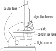
Many parts of the cell are hard to see under microscopes because they are colorless. In order to view them, scientists sometimes employ stains that mark various cell parts differently. One alternative to staining is a technique called phase contrast microscopy, which uses filters to emphasize the contrast between different parts of the cell.
The Electron Microscope
At high magnifications, light microscopes produce blurry images. In the 1950s, scientists invented a new type of microscope called the electron microscope, which offers increased image clarity, or resolving power. Electron microscopes are powerful enough to resolve individual fats and proteins. Light microscopes are still widely used, however, because electron microscopes are expensive and can only be used to view matter that is not living.
div>
Next to display next topic in the chapter.
Practice Questions
Test Prep Lessons With Video Lessons and Explained MCQ
Large number of solved practice MCQ with explanations. Video Lessons and 10 Fully explained Grand/Full Tests.